What Is a Prenatal Ultrasound?
A prenatal ultrasound is a safe and painless test that uses sound waves to make images that show the baby’s shape and position. It can be done in the first, second, or third trimester, depending on what the health care provider is looking for.
The American College of Obstetrics and Gynecologists (ACOG) recommends at least one ultrasound during a pregnancy, but many women have two or more. Women with high-risk pregnancies might have multiple ultrasounds during their pregnancy.
Why Are Prenatal Ultrasounds Done?
Ultrasounds were once used only in high-risk pregnancies, but have become so common that they’re often part of routine prenatal care.
During an ultrasound, sound waves are bounced off the baby’s bones and tissues to make an image showing the baby’s shape and position in the uterus. Also called a sonogram, sonograph, echogram, or ultrasonogram, an ultrasound is done to:
- confirm the expected date of delivery
- detect pregnancies outside the uterus
- see whether there might be more than one fetus
- see whether the fetus is growing at a normal rate
- record fetal heartbeat or breathing movements
- check the amount of amniotic fluid in the uterus
- check the position of the placenta (which can sometimes block the baby’s way out of the uterus) in late pregnancy
- guide doctors during other tests, like amniocentesis
- find structural defects that may indicate Down syndrome, spina bifida, or anencephaly (when the skull, scalp, and brain do not form properly)
- find other problems, such as congenital heart defects, cleft lip or palate, and gastrointestinal or kidney problems
What Happens During a Prenatal Ultrasound?
To prepare for a prenatal ultrasound, you might be asked to drink lots of fluids or not pee for a while so that your bladder will be full for the exam.
You’ll change into a cloth gown and lie on a table. The room is usually dark so the images can be seen clearly on the computer screen. A technician (sonographer) trained in ultrasound imaging will spread a clear gel on your abdomen. This gel helps with the transmission of the sound waves.
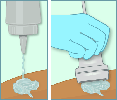
The technician will move a small wand (a transducer) over the gel. The transducer emits high-frequency sound waves and a computer measures how the sound waves bounce back from the body. The computer changes those sound waves into images. Sometimes a doctor will come in at the end of the test to meet with you and take a few more pictures.
The prenatal ultrasound is painless. You may feel a slight pressure on the belly as the transducer is moved over the body, and the gel may feel wet or cold. The test usually takes less than 30 minutes.
Sometimes an ultrasound test can be done through the vagina (called a transvaginal ultrasound) early in the pregnancy when the uterus and ovaries are better seen from that angle.
Some ultrasounds can show the fetus in three dimensions, like a photograph (a 3D ultrasound), and sometimes show movement (a 4D ultrasound). Doctors might use these to look for birth defects.
Should I Have a Prenatal Ultrasound?
This test is considered safe, but it’s up to you whether to have it. Talk to your health care provider to find out why and when this test is recommended for you.
When Are Prenatal Ultrasounds Done?
The timing of the test will depend on why the health provider recommends it.
A prenatal ultrasound can be done early in the first trimester to confirm and date the pregnancy, or during weeks 11–14 as part of the first trimester screening test.
Second-trimester ultrasounds (also called standard or “level 2” ultrasounds) usually are done between 18–20 weeks to examine the fetal anatomy and to confirm normal development. This test can often show the gender of the fetus (as long as the fetus is “cooperating” and in the right position). If you want your baby’s gender to be a surprise, make sure to tell the doctor or technician at the start of this test.
Third-trimester ultrasounds can examine the growth of the baby, the level of amniotic fluid, the position of the placenta, and the position of the fetus. Sometimes an ultrasound is part of a test called a biophysical profile (BPP) to see whether the fetus is getting enough oxygen. The BPP examines the baby’s breathing, movement, amount of amniotic fluid, tone, and heart rate response.
When Are the Results Available?
The technician can see the images right away, but a full evaluation may take up to 1 week if a doctor or specialist is not on site during the exam.
Depending on where you have the ultrasound done, the technician may be able to tell you that day if everything looks OK. But most radiology centers or health care providers prefer that technicians not comment until a specialist takes a look, even when everything is fine.